What is a DXA scan, and do you need one for bone health?
It's a test that comes up more and more in conversations about bone health: the DXA scan. But what exactly is it, and who actually needs one? As fitness and longevity content has proliferated on social media, this brief imaging test has quickly found its way into everyday health vocabulary.
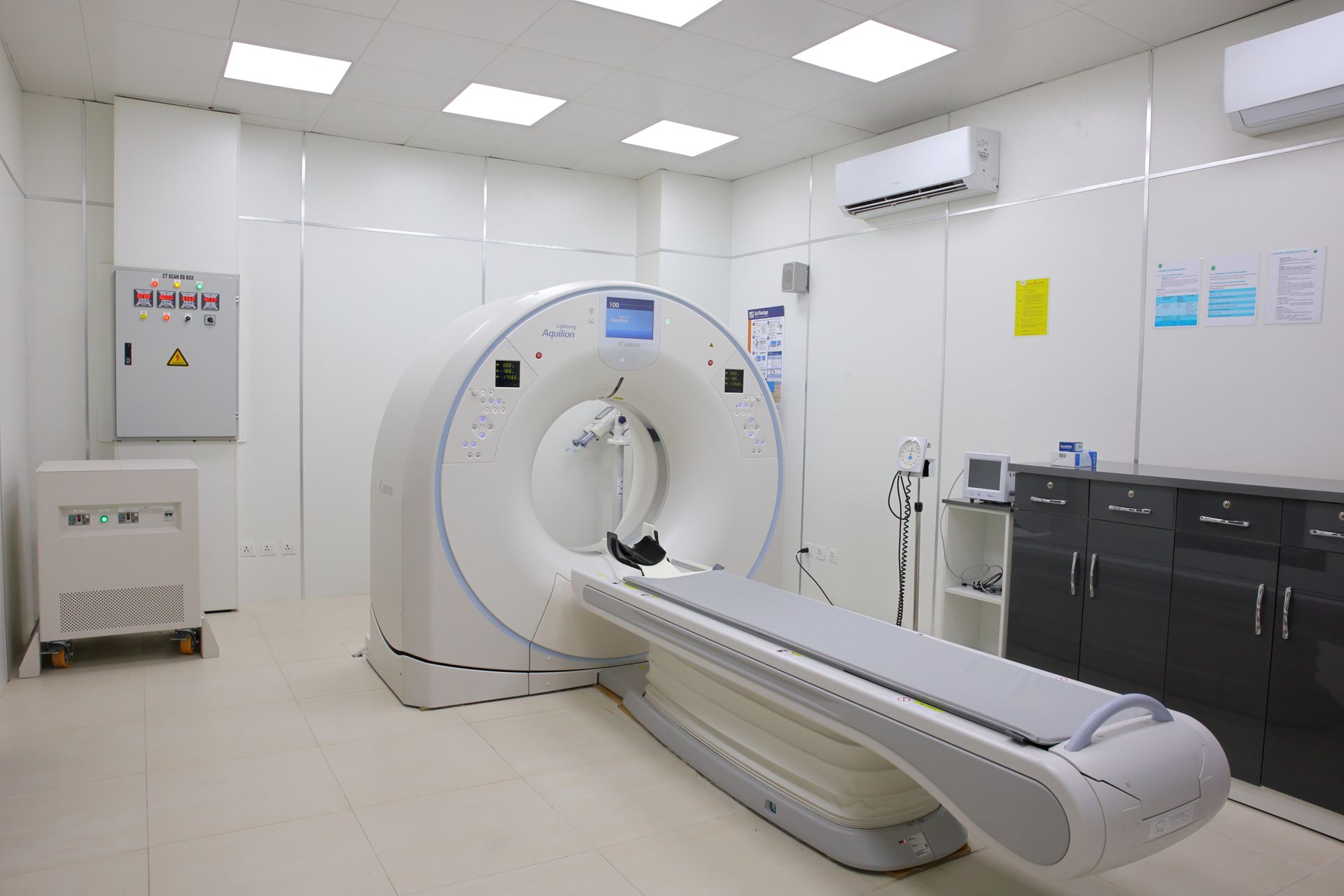
DXA — dual-energy X-ray absorptiometry — is an imaging test that uses low-dose X-rays to measure the strength and mineral content of bones. Dr Michael Perry, associate professor of radiology and medical imaging at the University of Virginia's school of medicine, describes it as "safe, quick and painless" and notes that patients need no special preparation beforehand.
The scan is typically taken at the lower spine, hip and sometimes the forearm — the areas most prone to fracture. Patients lie fully clothed on a table, and the entire process takes only a few minutes; it requires no needles, no contrast dye and no special diet, making it considerably more practical than many other imaging methods.
Results compare a person's bone density to that of an average young adult (the T-score) and to people of the same age (the Z-score). A low T-score can signal osteopenia or osteoporosis, conditions in which bones weaken and become more prone to fracture; these scores serve as the primary reference point doctors use to guide treatment decisions.
DXA scans are most often recommended for women over 65 and for men over 70 with certain risk factors. Early menopause, long-term steroid use, a family history of osteoporosis or low body weight can all be reasons to get scanned at a younger age; certain chronic conditions and extended use of specific medications can also move the recommended screening age earlier.
In recent years, the test has also gained a following among a different crowd: younger athletes and fitness enthusiasts interested in body composition. DXA can distinguish fat mass from lean muscle mass alongside bone density, which has made it appealing for tracking athletic performance; some professional athletes and elite sports clubs have begun incorporating regular DXA scans into their in-season monitoring routines.
Experts stress that not everyone needs the test. For a young, healthy adult with no risk factors, DXA offers limited clinical value — its usefulness is greatest for people whose fracture risk is genuinely elevated. Unnecessary screening can add cost to the health system and create needless anxiety for patients.
The radiation dose involved is extremely low — less than a standard chest X-ray — making the scan a safe option. Even so, results should be interpreted by a doctor alongside a patient's age, sex and medical history; a single number rarely offers a complete clinical picture on its own.
Osteoporosis often progresses silently, with the first sign frequently being an unexpected fracture. That's why regular DXA scanning for at-risk groups is considered a valuable tool for early intervention and treatment planning — catching the condition early allows medication and lifestyle changes to meaningfully lower fracture risk.
In short, DXA offers a quick, low-risk way to assess bone health — but who should get one, and when, depends on individual risk. Experts recommend anyone considering the test speak with a doctor first.
Read next

What is the Galleri test, and can a single blood draw really detect dozens of cancers?
Grail's Galleri test promises to screen a single blood sample for early signals of dozens of cancer types. As a US FDA advisory panel weighs the test, scientists caution that early evidence is promising but not yet conclusive.

How much strength training do you really need for a longer life?
A new study finds that just 90 to 120 minutes of strength training a week is linked to a 13% lower risk of early death, with the reduction climbing to around 45% when paired with regular aerobic exercise. The findings point to a clear "sweet spot" for longevity.

We know sleep, exercise and diet are vital for health. But why?
Sleep, exercise and diet have been treated for decades as separate prescriptions. New neuroscience research suggests the three habits produce remarkably similar effects on the brain and body, converging on a shared cellular mechanism.

Eli Lilly's new weight-loss pill Foundayo approved for use in the UK
Foundayo, an oral weight-loss pill made by Eli Lilly — the drugmaker behind the Mounjaro injection — has been approved for use in the UK. The pill offers a needle-free alternative that could widen access to obesity treatment.
England on track to become one of the first countries to eliminate hepatitis C
England is on track to become one of the first countries in the world to eliminate hepatitis C, a liver-damaging virus, health officials say. Widescale testing and modern antiviral treatment have driven down new infections and deaths over the past decade.